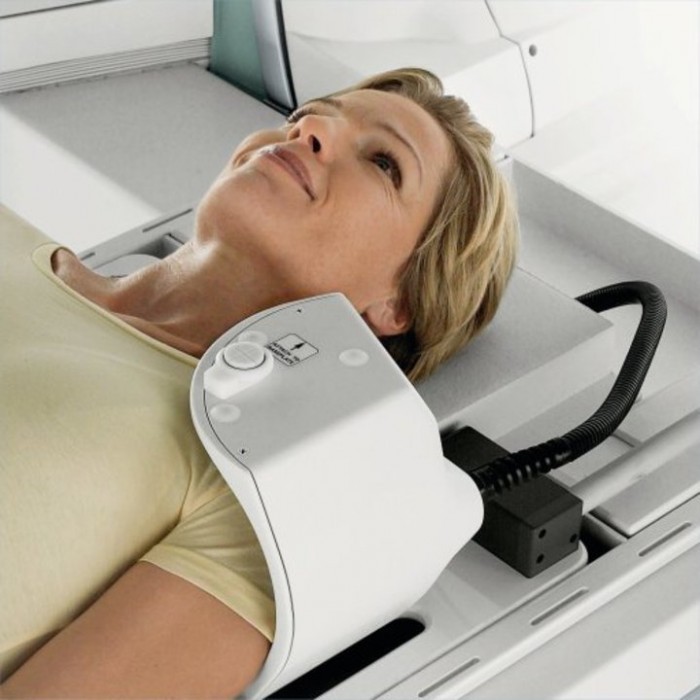
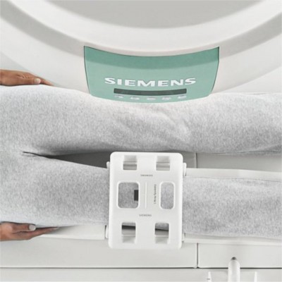
Orthopedic/Musculoskeletal MRI is used to examine bones, joints and soft tissues such as cartilage, tendons, and muscles for the presence of structural damage, defects, infection, etc. Metro provides musculoskeletal specialists.
Examples of Orthopedic/Musculoskeletal MRI are:
Each exam is tailored to our individual patients. Selection of the correct coil and protocol is done prior to your exam. Your doctor may recommend seeing a Musculoskeletal MRI specialist.
On most T1-weighted images, tissues with short T1 times (like subcutaneous fat or fatty bone marrow) appear bright; tissues with long T1 times (like fluid) appear dark. Solids (like cortical bone) also appear dark. If “fat saturation” is used, fat will appear dark on a T1-weighted image. The variation in sequences within each protocol allows the radiologist to identify any abnormality.
Each of our locations has a variety of coils specifically used for the body part that was ordered, and protocols that were approved by a radiologist.
It is important for your exam to be interpreted by a board certified radiologist.
Radiologists are medical doctors (MDs) who specialize in diagnosing and treating diseases and injuries using medical imaging techniques, such as x-rays, computed tomography (CT), magnetic resonance imaging (MRI), nuclear medicine, positron emission tomography (PET) and ultrasound.
Radiologists graduate from accredited medical schools, pass a licensing examination, and then go on to complete a residency of at least four years of unique post-graduate medical education in, among other topics:
These physicians complete a fellowship — one to two additional years of specialized training in a particular subspecialty of radiology, such as breast imaging, cardiovascular radiology or nuclear medicine.
The radiologists from Advanced Radiology, SC are board certified by the American Board of; an indication of a high level of training, and demonstrated excellence in the field.
Radiological procedures are medically prescribed and should only be conducted by appropriately trained and certified physicians under medically necessary circumstances. Radiologist physicians have four to six years of unique, specific, post–medical school training that includes radiation safety and ensure the optimal performance of radiological procedures and interpretation of medical images.
When you have a diagnostic imaging test, it is recommended to have a radiologist interpret your exam.
An arthrogram is a diagnostic test that examines the inside of a joint. It is done in two parts.
The test is done by first injecting contrast medium directly into the joint. This is usually done using fluoroscopy (a moving x-ray) to guide the placement of the needle to assuring that the contrast is in the correct location. The contrast will help differentiate the soft tissue structures, ligaments and tendons.
You will then go to an MRI scanner, where precise MR images are obtained.
Most common arthrograms are of the shoulder, because of the complexity of the joint. These are called MRI shoulder arthrograms or Shoulder MRI’s.